2 - Copy

Case Report:-

Bilaterally Occurring 4-Rooted Maxillary Second Molars : A Rare Care Report

Abstract

Knowledge of both normal and abnormal anatomy of the root canal system (extra canals and extra roots) dictates the parameters for execution of root canal therapy and can directly affect the outcome of the endodontic therapy. Maxillary second molars with two palatal roots, bilaterally is a rare dental anamoly. The diagnosis and treatment of extra root may create challenge for clinicians. The article also highlights the variations in the canal configuration and morphology with the methods to successfully diagnose, negotiate and treat the teeth with different anatomical variations. The case report underlines the importance of complete knowledge about root canal morphology which achieved by careful clinical and radiographic examination. The present case report describes root canal treatment of maxillary second molar with two palatal roots. Anatomic variations can occur in many teeth, and palatal root of maxillary second molar is not exception; so, the clinician should not focus only on the variations of mesio buccal root in the maxillary molars and careful radiographic examination and clinical evaluation of pulp chamber should be considered.

Key words: Root Canal Morphology, Palatal Root, Maxillary Second Molar, Anatomical Variations, Endodontic Treatment

Introduction

A thorough knowledge of tooth morphology, careful interpretation of angled radiographs, proper access preparation and a detailed exploration of the interior of the tooth are essential prerequisites for a successful treatment outcome. Cleaning, shaping and obturation of the entire root canal system “three dimensionally” is important and- essential for successful endodontic therapy. Magnification and illumination are aids that must be utilized’ to achieve this goal.

The main reasons endodontic failure are apical percolation and microorganisms persisting in the pulp space because of incomplete instrumentation inadequate cleaning insufficient canal obturation and presence of intreated canal.  Treatment may be unsuccessful because dentist fails to recognize the unusual canal configuration. Therefore, a thorough knowledge of root anatomy, root canal morphology and a good anticipation of their possible morphologic variations are essential and will help to reduce endodontic failure. Literature review demonstrates an extensive number of variations pertaining to the form, configuration and the number of roots and root canals present in the maxillary molars.

Pecora et al evaluated the anatomy of 370 maxillary molars and found that the maxillary first, second and third molars showed three canals in 75%, 58% and 68% of the teeth, respectively. Four canals were found in 61.1% of maxillary first molars, in 42% of second molars, and in 32% of third molars. The fourth canal was mainly found in the mesiobuccal root of the teeth. In the assessment of root canal configuration of maxillary first permanent molars in an Iranian population performed by Shahi et al, 58.4% of maxillary first molars demonstrated four root canals and in 0.73% of studied first molars had two palatal root canals. It had been showed that the patient’s age was an important predictor of the detection of fewer canals in maxillary molars. This is likely because of the calcification and morphologic changes that occur with age and makes discovery of canals root number of maximum difficult. This might be one of the reasons of the big discrepancies in the number of detected second, mesio buccal canals in different studies. Corcoran et al stated that, operator experience has a positive effect on the number of canals located in maxillary molars’.

This case report may intensify the complexity of maxillary molar variation and is intended to reinforce clinician’s awareness of rare morphology of root canals. The authors review related literature, describes the clinical cases and make considerations about how dentists could perform the treatment of maxillary molars to achieve successful results in similar cases.

Case Report

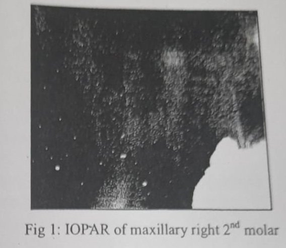
A 32years old female with a non- contributory medical history was referred to the Department of Conservative Dentistry and Endodontics, because of pain and percussion tenderness on the maxillary right second examination showed the molar. Clinical extensive carious lesion & heavily restored maxillary right first molar. Second molar revealed severe tenderness to percussion. There was no response to heat and cold test on both first and second molar. The preoperative radiograph showed root canal filling in first molar & an extensive carious lesion at mesial surface, which possibly accounted for the inflammatory process together with the pain reported by the patient in maxillary second molar (Fig 1).

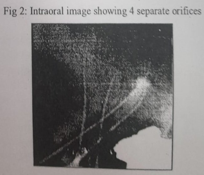
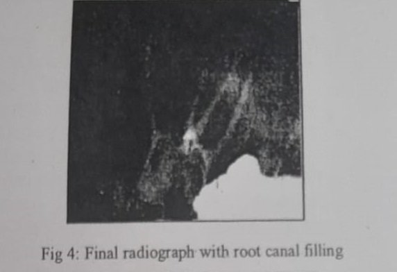
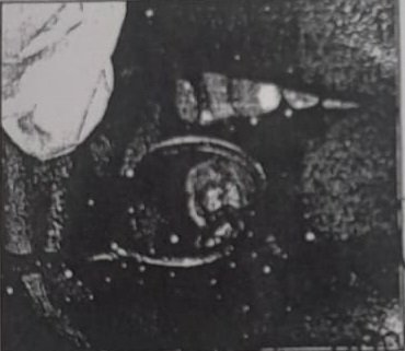
The maxillary right second molar was prepared for nonsurgical root canal treatment. The patient received local anesthesia of 2% Lidocaine with 1:100000 epinephrine (LA BRAND). The caries was removed and access preparation done. With a triangular access 3 orifices of mesio-buccal, disto-buccal, palatal were located. But the palatal canal, initially found was positioned slight distal to the usual anatomic position. The working lengths were determined. Radiograph taken with tube shift technique due to unusual anatomic location of palatal canal orifice, revealed an extra palatal root which was mesially placed. Ultrasonic tips (Dentsply Maillefer, Ballaigues, Switzerland) were used to remove the dentin & DG-16 Endodontic explorer used to locate the canal orifice (Fig 2). The working lengths were determined with apex locater (Propex II, and Switzerland) Dentsply Maillefer, controlled radiographically (Fig 3). The cleaning and shaping of canals were done by passive Crown Down technique with HyFlex® CMTM NiTi Rotary files (0.04 taper 25 no. files, Coltène/Whaledent). EDTA (dentsply maillefer glyde syringe kit) was used to lubricate canal. Canals were irri,ated with 6% during sodium hypochlorite instrumentation. After completion of canal preparation, pulp chamber was sealed with Cavit (ESPE, Seefeld, Germany) and patient was recalled next day for root canal filling. Next day patient was found asymptomatic and the canal were obturated with cold lateral condensation technique with size 25, 0.04% taper gutta- percha cones ( Coltene/Whaledent) and AH plus sealer (Dentsply Maillerfer) permanent restoration was done (Fig 4 and 5).


Discussion

Anatomical variations can occur in maxillary permanent molars. Christie et al14 speculated that maxillary second molars with two palatal roots may be encountered once every three years in a busy endodontic practice. The study conducted by Libfield and Rotstein showed that the incidence of two palatal roots in maxillary second molars was only 0.4%. Peikoff et al observed that 1.4% of maxillary second molars may have second palatal root.

The second maxillary molar usually has one canal in each root, however; it may have two or three mesiobuccal canals, one or two distobuccal canals or two palatal canals. It has been reported that second maxillary molar show two canals in the mesiobuccal roots in up to 58% of the cases’. The frequency of reports on two palatal roots in second maxillary molar is low Slowey first reported maxillary second molar with two palatal roots”. Thews et al also reported two maxillary second molar with this anatomic variation. Stone and Stroner examined more than 500 extracted molars and found less than 2% incidence of more than one palatal canal13. Christie et all showed that the highest occurrence of two palatal canals in double palatal roots was found in the maxillary second molar.

In this case radiograph of contra lateral maxillary left second molar was also found with 4 roots including 2 palatal roots. The present case report describes case of maxillary second molar with a canal configuration rarely reported in the literature. These teeth had four roots with four root canals, mesio-buccal root with single canal, two individual palatal roots (mesio-palatal and disto-palatal) with its own separate canal and disto-buccal root with a single canal.

When examining the pre-operative periapical radiographs of maxillary molars, if the outline of roots are unclear, if the root canals show sharp density changes or if the apices cannot be well defined, then extra roots can be suspected. Horizontally angled radiographs can be helpful to distinguish the multiple root morphology of molars. Sometimes the presence of palatogingival groove also indicates the presence of two palatal roots because this represents an abortive attempt to form two palatal roots. However, the most definitive means of determining root canal morphology is visualizing the pattern of the pulp chamber floor.

Gulabivala et al, have classified four rooted maxillary molars into three types: Type I with long tortuous and divergent palatal roots, Type II with short, blunt and parallel roots, Type III with convergent roots and Type IV – distinctly divergent fourth distobuccal root. The tooth described here had totally separated palatal roots, each with a distinct root canal.

Conclusion

The root canal anatomy of each tooth in the human dentition has certain common characteristics as well as numerous atypical ones that can be a roadmap to successful endodontics. Knowledge of the existence of these variations is important for both diagnostic and treatment standpoints. Thus it is essential to highlight the need to look for unusual morphology and additional roots and root canals so as to achieve a good endodontic outcome.

References

1. Vertucci FJ. Root canal morphology and its relationship to endodontic procedures. Endod Topics. 2005; 10. 3-29.

2. Harry L, Ilan R. Incidence of four rooted maxillary second molar: Literature review and radiographic survey of 1200 teeth. J Endod 1989; 15: 129-31.

3. Siqueria JF, Rocas IN. Clinical implications and microbiology of bacterial persistence after treatment procedures. J Endod 2008; 34: 1291-301.

4. Pecora JD, Woelfel JB, Sousa Neto MD, Issa EP. Morphologic study of the maxillary molars. Part ii: internal anatomy. Braz Dent J. 1992; 3: 53-57.

 5. Shahi S, Yavari HR, Rahimi S, Ahmadi A. Root canal configuration of maxillary first permanent molars in an Iranian population. J Dent Res Dent Clin Dent Prospect. 2007; 1: 1-5.

6. Iqbal M, fillmore E. Preoperative predictor of the canal clinically deteced in maxillary molars: a pennendo database study. J Endod. 2008; 34: 413-16.

7. Corcoran J, Apicella MJ, Mines P. The effect of operator experience in locating additional canals in maxillary molars. J Endod. 2007; 33: 15-17.

8. Libfeld H, Rotstein I. Incidence o. four rooted maxillary second molars: literature review and radiographic survey of 1200 teeth. J Endod. 1989; 15: 129-31.

9. Shalabi RM, Omer OE, Glennon J. Root canal anatomy of maxillary first and second permanent molars. Int Endod J. 2000; 33: 405-14.

10. Hülsmann H. A maxillary first molar with two disto-buccal root canals. J Endod. 1997; 23: 707-8.

11. Slowey RR. Radiographic aids in the detection of extra root canals. Oral surg oral med oral pathol oral radiol endod. 1974; 37: 762-72.

12. Thews ME, Kemp WB, Jones CR. Aberrations in palatal root and root canal morphology of two maxillary first molars. J Endod. 1979; 5: 94-6.

13. Stone LH, Stroner WF. Maxillary molars demonstrating more than one palatal root canal. Oral surg oral med oral pathol. 1981; 51: 649-52.

14. Christie WH, Peikoff MD, Fogel HM. Maxillary molars with two palatal roots: a retrospective clinical study. J Endod. 1991; 17: 80-4.

Acknowledgement

We wish to thank Dr Krishna Prasad (MDS) (Professor and Head, Department of Conservative Dentistry and Endodontics, Sharad Pawar Dental College, Data Megha Institute of Medical Sciences, Sawangi (M), Wardha (Maharasta) for his constant Support, Motivation and Encouragement during the Preparation of this Manuscript.

Corresponding author:

Dr Deepak Raisingani, Professor, Department of Conservative Dentistry and Endodontics, Mahatma Gandhi Dental College And Hospital, Mahatma Gandhi University Of Medical Sciences and Technology, Sitapura, Jaipur-302022, Rajasthan. +91 9414221345 draisingani@gmail.com.

Effect of Methods of Irrigation and Different Irrigant Combination on Root Canal Debris Removal in Curved and Straight Canal – An in Vitro Study.

ABSTRACT
Background-Although during root canal therapy we rely primarily on instrumentation for canal debridement, irrigation is an important adjunct. It facilitates cleaning of root canal system by flushing debris as well as serving as a bactericidal agent: Presence of debris on prepared root canal surface prevents efficient removal of microorganisms & completes sealing of root canal walls which is one of the major goal to be achieved. Aim and methodology-The aim of this in vitro study is to evaluate the debris removal efficacy of combination 5% NaOCl +17% EDTA and 5% NaOCl +1% tetracycline hydrochloride irrigants when they were used in conjunction with plain and Navi Tip Fx Needle in straight and curved canals. As the type of irrigants, the irrigation delivery system and canal curvature are the determining factors for the root canal cleanliness, this study has been undertaken to assess which of these factors are crucial for success of the root canal treatment. Statistical analysis used- Mean, mann-whitney U test, Chi Square test. Result and conclusion- Navi Tip Fx is better in debris removal compared with plain needle but efficacy of Navi Tip Fx is reduced in apical one third of canals in both straight and curved canals. 5% Sodium hypochlorite +17% EDTA irrigants combination is more efficient in total debris removal compared with 5% sodium hypochlorite+1% tetracycline hydrochloride. Keywords- Navi Tip Fx, Tetracycline hydrochloride, EDTA, NaOCl

INTRODUCTION

Successful endodontic therapy involves a series of procedures that begins with adequate knowledge of the biology of the pulp and periapical tissues, and ends with the subsequent evaluation of the treatment rendered. Cleaning and shaping of the root canal is one of the most important and demanding aspect of this treatment. Debridement of the root canal system is essential for endodontic success, with irrigation being a vital part of root canal debridement. According to Cohen “what is taken out of the root canal may be more important than what is put into the canal”. Ideally, root canal irrigants should flush out debris, dissolve organic tissue, kill microbes, destroy microbial byproducts An Excerpt from the Dissertation submitted to Pt. B.D. Sharma University of Health Sciences, Rohtak, Year- 2009

and remove smear layer. The purpose of irrigation is twofold: to remove gross debris originating from pulp tissue; possible bacteria; the organic component and to remove smear layer; consisting mainly of the inorganic component.Heredia et al stated that the most widely used irrigant for root canal treatment is sodium hypochlorite (NaOCl) at concentration of 0.5% to 5.25%. The tissue dissolving capacity and microbial activity of NaOCl makes it an excellent irrigating solution, but it has only limited effect on the dissolution of smear layer. Acid solutions have been recommended for removing the smear layer including sodium salt of ethylenediaminetetracetic acid (EDTA) which is most active at a concentration of 15 to 17% and pH of 7 to 8.Furthermore, after instrumentation, a smear layer is formed that is a surface film of 1-2μm thickness that remains adherent to the root canal wall. This smear layer consists of dentin particles, pulp tissue, bacterial components, as well as retained irrigants and it occludes the dentinal tubular openings. Al Hadlaq M et al reported that the chemo mechanical action of sodium hypochlorite removes loosely attached debris or organic material; whereas chelating agents are required to remove the smear layer. Barkhordar et al reported that the usefulness of tetracycline is attributed to its antibacterial activity and its recently discovered ability to inhibit mammalian collagenases. Haznedaroglu F et al showed that tetracycline hydrochloride (HCl), an acidic solution with bacteriostatic properties, has been considered to be suitable for endodontic irrigation’. According to Ingle irrigation delivery system is also one of the important part for action of root canal irrigants. Irrigation is commonly applied by a syringe and a needle’. A Recently introduced irrigation needle (Navi Tip Fx Ultradent) which is covered with brush is used in this study. Gunday et al stated that in addition to the canal instruments and preparation techniques used, root canal morphology and degree of curvature are also the determining factors during the root canal preparation’. In curved canal the deformation of canal instruments places stress on the instrument, with tensile stresses exerted on the noncurved parts and compressive stresses from the curved parts of the canal instruments. When the curvature of canal increases distorted part of the file becomes greater and the risk of breakage increases. The aim of this in vitro study is to evaluate the debris removal efficacy of combination 5% NaOCI + 17% EDTA and 5% NaOCl + 1% tetracycline hydrochloride irrigants when they were used in conjunction with plain and Navi Tip Ex Needle in straight and curved canals. As the type of irrigants, the irrigation delivery system and canal curvature are the determining factors for the root canal cleanliness, this study has been undertaken to assess which of these factors are crucial for success of the root canal treatment.

Methodology

In this in vitro study total hundred single rooted (50 straight and 50 curved) noncarious, extracted, human teeth were collected. Exterior surface of all teeth were thoroughly cleaned of organic debris and calculus with ultrasonic scaler. Among the curved root the canal curvature was selected between 10-20 degree. Canal curvature was measured as described by Schneider (1971) from preoperative radiograph with the help of RVG (GENDEX, DENTSPLY). The teeth were stored in saline till use.

All the teeth were sectioned at cementoenamel junction with diamond disc in presence of water. Patency of the canal was checked by inserting a size 10 K-file (Mani) into the canal so that the tip of the file was just visible through the apical foramen. Individual working length was calculated 0.5mm short from this position. Samples were randomly divided into 10 groups, (8 experimental and 2 control groups) in this study. Number of samples were kept 10 in each experimental and control groups. All the Samples in experimental group were prepared with K file (up to #40) by step back technique as described by Grossman using different irrigants.

Experimental samples were divided into following 8 groups on the basis of irrigating solutions, canal curvature and method of irrigation procedure.

> Group IA: Samples with straight canals were irrigated with plain needle using 1 ml of 5% NaOCI followed by 1 ml of 17% EDTA solution alternatively throughout the preparation.

> Group IIA: Samples with straight canals were irrigated with Navi Tip FX using 1 ml of 5% NaOCl fellowed by 1 ml of 17% EDTA solution alternatively throughout the preparation.

> Group IIIA: Samples with straight canals were irrigated with plain needle using 1 ml of 5% NaOCl followed by 1 ml of 1%. tetracycline hydrochloride solution alternatively throughout the preparation.

> Group IVA: Samples with straight canals were irrigated with Navi Tip FX using 1 ml of 5% NaOCl followed by 1 ml of 1% tetracycline hydrochloride solution alternatively throughout the preparation.

> Group IB: Samples with curved canals were irrigated with plain needle using 1 ml of 5% NaOCl followed by 1 ml of 17% EDTA solution alternatively throughout the preparation.

> Group IIB-Samples with curved canals were irrigated with Navi Tip FX using 1 ml of 5% NaOCI followed by 1 ml of 17% EDTA solution alternatively throughout the preparation.

> Group IIIB-Samples with curved canals were irrigated with plain needle using 1 ml of 5% NaOCI followed by 1 ml of 1% tetracycline hydrochloride solution alternatively throughout the preparation.

> Group IVB: Samples with curved canals were irrigated with Navi Tip FX using 1 ml of 5% NaOCl followed by 1 ml of 1% tetracycline hydrochloride solution alternatively throughout the preparation.

In control groups no irrigation and instrumentation was performed.

> Group VA Samples with straight canals which were not instrumented.

> Group VB Samples with curved canals which were not instrumented.

During the cleaning and shaping each sample was irrigated with 1 ml of irrigant after each change of instrument. Distilled water was used in between two irrigation as well for final rinse to neutralize the effect of these irrigants. Vertical grooves along the long axis of Samples were made on all the samples with the help of diamond disc in presence of moisture. Then all the samples were split into two halves using chisel and mallet. The split samples were prepared for SEM analysis.

Canal cleanliness evaluation

Root canal debris is defined as dentine chips, pulpal remnants and particles loosely attached to the canal wall. Scoring, of debris was performed at 500x magnification

Scoring system for debris as given by Peters OA et al

> Score 1: Clean root canal wall, only few small debris particles.

> Score2: Few small agglomerations of debris covering less than 25% of canal wall area.

> Score 3: Many agglomerations of debris covering 25% to 50% of root canal wall area.

> Score 4: 50% to 75% of the root canal wall area covered by debris.

> Score 5: More than 75% of root canal wall area covered by debris.

Statistical analysis used- Mean, Mann- Whitney U test, Chi-square test. Statistical software used statistical package for social sciences (SPSS). version 12.0.

Significance at different levels among all groups for debris removal analyzed by chi-square

test.

Mann-Whitney U test-it is a nonparametric test for assessing whether two independent samples of observations come from the same distribution.

Statistical comparison for straight vs curved canals, plain vs Navi Tip FX and irrigating solutions (5% NaOCl + 17% EDTA vs 5% NaOCl +1% tetracycline hydrochloride) by Mann-Whitney test.

Results and observations

After preparation all samples were viewed under SEM and photomicrographs were taken at 500X. Scoring was done for all samples according to scoring system given by Peters OA et al. All samples were evaluated and scored at cervical, middle and apical level.

Table – 1

Comparative evaluation of mean debris score of different goups at different levels

CervicalMiddleApicalP value
Group lA2.02.43.6.000
Group llA1.82.03.4.000
Group lllA2.42.84.0.000
Group lVA2.62.24.6.000
Group lB1.83.04.2.000
Group llB2.22.42.8.000
Group lllB2.23.24.6.000
Group lVB1.82.44.8.000
Group VA5.05.05.0.000
Group VB5.05.05.0.000

Table -2

Showing mean, SD and p Values at different levels of tooth

DISCUSSION

The primary function of an endodontic irrigant is to remove debris loosened but not removed by instrumentation. Moreover, some irrigants have property to remove the smear layer. Hulsmann et al found that the smear layer consist of inorganic and organic substances including fragments of odontoblastic processes, micro-organisms and necrotic material”. It is made up of a superficial layer on the canal walls approximately 1-2pm thick and a deeper layer packed into the dentinal tubules up to 40μm. Degree of smear layer removal depends upon the type of irrigants used. In this study the mean debris score for control and experimental group were 15.0 and 8.65 respectively. The uninstrumented canals showed walls completely covered with tissue confirming the finding of Heard F et al that specimen preparation alone did not remove tissue from canal walls”. The mean debris score for straight and curved canals were 2.82 and 2.95 respectively. While at cervical, middle and apical level the mean debris scores were 2.2, 2.25, 3.9 for straight canals and 2.0, 2.75, 4.10 for curved canals. These results are similar as obtained by Ciucchi et al, Garberoglio et al, and Teixeira et al 121314. This may be attributed to the fact that as reported by Ciucchi et al, limited distribution of the irrigants is because of limited size and pronounced curvature of the canal which are obstacles to the optimal apical flooding of irrigants”. Moreover Garberoglio et al stated that a major challenge in

clinical endodontics is the preparation of apical region of the root canal”. The access is often difficult because of its anatomy resulting in reducing effects of the irrigants at the level of 1 to 3mm from the apex. In these areas irrigants are not effective in cleaning the root canal system and use of powerful irrigating agent is contraindicated because of the risk of injury to periradicular tissues In this study the mean debris score for samples irrigated with 5% NaOCl + 17% EDTA group was 2.63 while for samples irrigated with 5% NaOCl + 1% tetracycline hydrochloride it was 3.13. Mann Whitney test showed that there was significant difference for debris removal in between the two irrigating solution group (p = .001). Barkhordar et al reported that irrigation of teeth with aqueous solutions of doxycycline HCl eliminated smear layer in concentration dependent manner. Sen et al reported that the removal of smear layer as well as soft tissue and debris can be expedited by the alternate use of EDTA and NaOC15. According to them 5.25% NaOCl and 17% EDTA concentration is most effective concentration in removing debris from root canal walls. In this study mean debris score for the samples in which plain needle used was 3.02 and for Navi Tip Fx, it was 2.75. It showed that Navi Tip FX is more efficient in debris removal .Mann Whitney analysis showed that difference of debris removal was statistically significant between both Needle groups (p=0.04). As suggested by Al Hadlaq et al that Navi Tip Fx (ultradent) which is a brush covered needle would produce cleaner coronal one third of instrumented root canal walls of single rooted teeth compared to brushless (Plain needle)”. However, the efficacy of the Navi Tip Fx in teeth with more complex root system such as mesial roots of upper and lower first molar has to be evaluated. More research is needed for the quest of irrigating solution and the irrigation delivery system that can fulfill the requirements of ideal irrigation system for increased success of root canal treatment.


CONCLUSION

Results of the study showed that:

> Total debris removal was better in straight canals compared with curved canals.

> Debris removal was less from apical one third in both straight and curved canals due to limited distribution of irrigants.

> Navi Tip Fx is better in debris removal compared with plain needle but efficacy of Navi Tip Fx is reduced in apical one third of canals in both straight and curved canals,

> 5% Sodium hypochlorite + 17% EDTA irrigants combination is more efficient in total debris removal compared with 5% sodium hypochlorite+1% tetracycline hydrochloride.

Within limitation of this in vitro study, it can be concluded that 5% Sodium hypochlorite + 17% EDTA irrigant combination was more efficient in total debris removal compared to 5% sodium hypochlorite +1% tetracycline hydrochloride. Moreover Navi Tip Fx was more efficient in debris removal from root canal than plain needle, however further research is needed on the use of Navi Tip Fx before its routine use in Endodontic practice.

BIBLIOGRAPHY

1. Peters OA, Peters CI. Cleaning and shaping of the root canal system. Cohen S, Hargreaves KM. Pathways of pulp. 9th ed. New Delhi: Elsevier; 2006.p.290-357

2. Nielsen BA, Baumgartner JC. Comparison of EndoVac System to needle irrigation of Root Canals. J Endod 2007; 33 (x): 1 – 5.

3. Heredia MP, Luque CMF, Roidriguez MPG. The effectiveness of different acid irrigating solutions in root canal cleaning after hand and rotary instrumentation. J Endod 2006;32:993-97.

4. Al-Hadlaq SM, Al-Turaiki SA, Al-Sulami U. Efficacy of a new brush-covered irrigation needle in removing root canal debris: A Scanning Electron Microscopic study. J Endod 2006; 32:1181 – 84.

5. Barkhordar RA, Watanabe LG, Marshall GW, Hussain MZ. Removals of intracanal smear by doxycycline in vitro. Oral Surg Oral Med Oral Pathol Oral Radiol Endod 1997; 84: 420-23

6. Haznedaroglue F and Ersev H. Tetracycline HCl Solution as a Root Canal Irrigant. J Endod 2001; 12(27): 738 – 40.

7. Peters OA, Koka RS. Preparation of coronal and radicular spaces. Ingle JI, Bakland LK, Baumgartner JC. Endodontics. 6th ed. Hamilton, Ontario. BC Decker ;2008.p:877-991.

8. Gunday M, Sazak H, Garip Y. A comparative study of three different root canal curvature measurement techniques and measuring the canal access angle in curved canals. J Endod 2005; 31 (11): 796 – 98.

9. Peters OA, Meddent, Barbakow F. Effect of irrigation on debris and smear layer on canal walls prepared by two rotary techniques: A SEM study. J Endod 200026(16-10

10. Hulsmann M, med dent, Rummelin C, Schäfers F. Root canal cleanliness after preparation with different endodontic hand pieces and hand instruments: A comparative SEM investigation. J Endod 1997; 23(5):301 – 6.

11. Heard F and Walton RE. Scanning electron microscope study comparing four root canal preparation techniques in small curved canals. Int Endod J 1997; 30: 323 – 31.

12. Ciucchi B, Khettabi M and Holz J. The effectiveness of different endodontic irrigation procedures on the removal of the smear layer: a scanning electron microscopic study. Int Endod J 1989; 22: 21-28.

13. Garberoglio R Becce C. Smear layer removal by root canal irrigants – A comparative scanning electron microscopic study. Oral Surg Oral Med Oral Pathol 1994; 78: 359-67.

14. Teixeira CS,Felippe MCS,Felippee WT.The effect of application time of EDTA and NaOCl on intracanal smear layer removal :an SEM analysis. Int Endod J 2005; 38:285-90.

15. Sen BH, Wesselink PR, Turkun M. The smear layer. a phenomenon in root canal therapy. Int Endod J 1995; 28:141-48.

Address for Correspondence

Dr. Medhavi Sharma House No. 494, Surya Nagar,

Gopalpura Bypass, Jaipur – 302015

E-mail: drmedhavisharma@gmail.com