Bilaterally Occurring 4-Rooted Maxillary Second Molars : A Rare Care Report
Abstract
Knowledge of both normal and abnormal anatomy of the root canal system (extra canals and extra roots) dictates the parameters for execution of root canal therapy and can directly affect the outcome of the endodontic therapy. Maxillary second molars with two palatal roots, bilaterally is a rare dental anamoly. The diagnosis and treatment of extra root may create challenge for clinicians. The article also highlights the variations in the canal configuration and morphology with the methods to successfully diagnose, negotiate and treat the teeth with different anatomical variations. The case report underlines the importance of complete knowledge about root canal morphology which achieved by careful clinical and radiographic examination. The present case report describes root canal treatment of maxillary second molar with two palatal roots. Anatomic variations can occur in many teeth, and palatal root of maxillary second molar is not exception; so, the clinician should not focus only on the variations of mesio buccal root in the maxillary molars and careful radiographic examination and clinical evaluation of pulp chamber should be considered.
Key words: Root Canal Morphology, Palatal Root, Maxillary Second Molar, Anatomical Variations, Endodontic Treatment
Introduction
A thorough knowledge of tooth morphology, careful interpretation of angled radiographs, proper access preparation and a detailed exploration of the interior of the tooth are essential prerequisites for a successful treatment outcome. Cleaning, shaping and obturation of the entire root canal system “three dimensionally” is important and- essential for successful endodontic therapy. Magnification and illumination are aids that must be utilized’ to achieve this goal.
The main reasons endodontic failure are apical percolation and microorganisms persisting in the pulp space because of incomplete instrumentation inadequate cleaning insufficient canal obturation and presence of intreated canal. Treatment may be unsuccessful because dentist fails to recognize the unusual canal configuration. Therefore, a thorough knowledge of root anatomy, root canal morphology and a good anticipation of their possible morphologic variations are essential and will help to reduce endodontic failure. Literature review demonstrates an extensive number of variations pertaining to the form, configuration and the number of roots and root canals present in the maxillary molars.
Pecora et al evaluated the anatomy of 370 maxillary molars and found that the maxillary first, second and third molars showed three canals in 75%, 58% and 68% of the teeth, respectively. Four canals were found in 61.1% of maxillary first molars, in 42% of second molars, and in 32% of third molars. The fourth canal was mainly found in the mesiobuccal root of the teeth. In the assessment of root canal configuration of maxillary first permanent molars in an Iranian population performed by Shahi et al, 58.4% of maxillary first molars demonstrated four root canals and in 0.73% of studied first molars had two palatal root canals. It had been showed that the patient’s age was an important predictor of the detection of fewer canals in maxillary molars. This is likely because of the calcification and morphologic changes that occur with age and makes discovery of canals root number of maximum difficult. This might be one of the reasons of the big discrepancies in the number of detected second, mesio buccal canals in different studies. Corcoran et al stated that, operator experience has a positive effect on the number of canals located in maxillary molars’.
This case report may intensify the complexity of maxillary molar variation and is intended to reinforce clinician’s awareness of rare morphology of root canals. The authors review related literature, describes the clinical cases and make considerations about how dentists could perform the treatment of maxillary molars to achieve successful results in similar cases.
Case Report
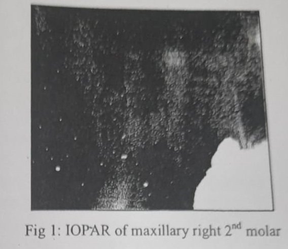
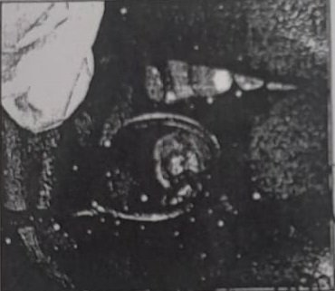
A 32years old female with a non- contributory medical history was referred to the Department of Conservative Dentistry and Endodontics, because of pain and percussion tenderness on the maxillary right second examination showed the molar. Clinical extensive carious lesion & heavily restored maxillary right first molar. Second molar revealed severe tenderness to percussion. There was no response to heat and cold test on both first and second molar. The preoperative radiograph showed root canal filling in first molar & an extensive carious lesion at mesial surface, which possibly accounted for the inflammatory process together with the pain reported by the patient in maxillary second molar (Fig 1).
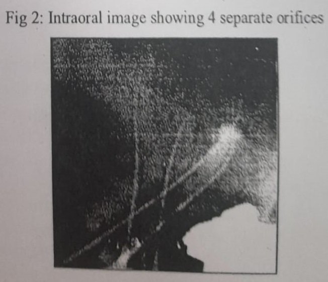
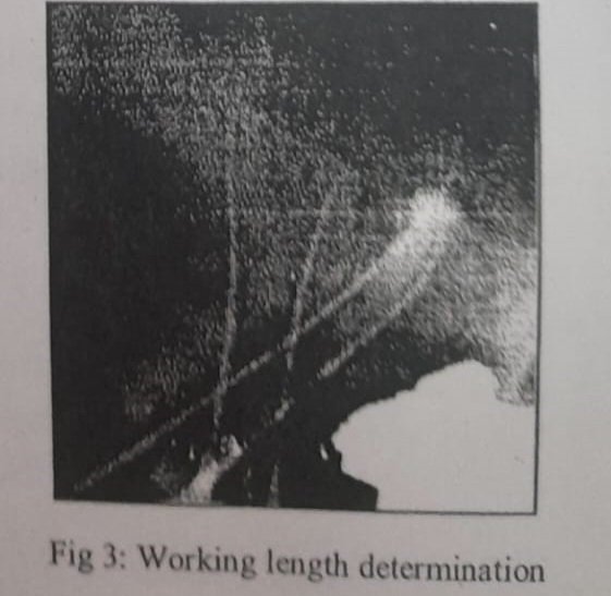
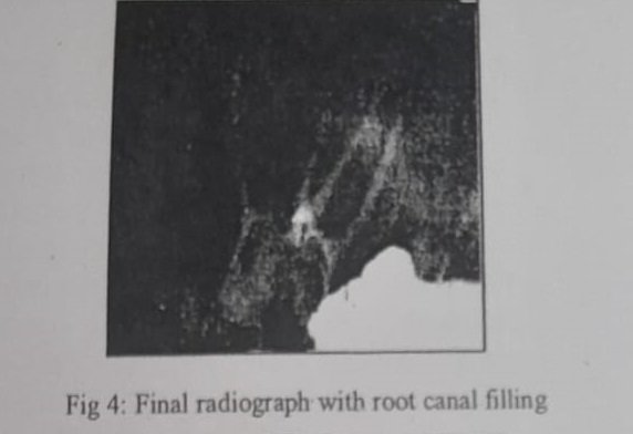
The maxillary right second molar was prepared for nonsurgical root canal treatment. The patient received local anesthesia of 2% Lidocaine with 1:100000 epinephrine (LA BRAND). The caries was removed and access preparation done. With a triangular access 3 orifices of mesio-buccal, disto-buccal, palatal were located. But the palatal canal, initially found was positioned slight distal to the usual anatomic position. The working lengths were determined. Radiograph taken with tube shift technique due to unusual anatomic location of palatal canal orifice, revealed an extra palatal root which was mesially placed. Ultrasonic tips (Dentsply Maillefer, Ballaigues, Switzerland) were used to remove the dentin & DG-16 Endodontic explorer used to locate the canal orifice (Fig 2). The working lengths were determined with apex locater (Propex II, and Switzerland) Dentsply Maillefer, controlled radiographically (Fig 3). The cleaning and shaping of canals were done by passive Crown Down technique with HyFlex® CMTM NiTi Rotary files (0.04 taper 25 no. files, Coltène/Whaledent). EDTA (dentsply maillefer glyde syringe kit) was used to lubricate canal. Canals were irri,ated with 6% during sodium hypochlorite instrumentation. After completion of canal preparation, pulp chamber was sealed with Cavit (ESPE, Seefeld, Germany) and patient was recalled next day for root canal filling. Next day patient was found asymptomatic and the canal were obturated with cold lateral condensation technique with size 25, 0.04% taper gutta- percha cones ( Coltene/Whaledent) and AH plus sealer (Dentsply Maillerfer) permanent restoration was done (Fig 4 and 5).
Discussion
Anatomical variations can occur in maxillary permanent molars. Christie et al14 speculated that maxillary second molars with two palatal roots may be encountered once every three years in a busy endodontic practice. The study conducted by Libfield and Rotstein showed that the incidence of two palatal roots in maxillary second molars was only 0.4%. Peikoff et al observed that 1.4% of maxillary second molars may have second palatal root.
The second maxillary molar usually has one canal in each root, however; it may have two or three mesiobuccal canals, one or two distobuccal canals or two palatal canals. It has been reported that second maxillary molar show two canals in the mesiobuccal roots in up to 58% of the cases’. The frequency of reports on two palatal roots in second maxillary molar is low Slowey first reported maxillary second molar with two palatal roots”. Thews et al also reported two maxillary second molar with this anatomic variation. Stone and Stroner examined more than 500 extracted molars and found less than 2% incidence of more than one palatal canal13. Christie et all showed that the highest occurrence of two palatal canals in double palatal roots was found in the maxillary second molar.
In this case radiograph of contra lateral maxillary left second molar was also found with 4 roots including 2 palatal roots. The present case report describes case of maxillary second molar with a canal configuration rarely reported in the literature. These teeth had four roots with four root canals, mesio-buccal root with single canal, two individual palatal roots (mesio-palatal and disto-palatal) with its own separate canal and disto-buccal root with a single canal.
When examining the pre-operative periapical radiographs of maxillary molars, if the outline of roots are unclear, if the root canals show sharp density changes or if the apices cannot be well defined, then extra roots can be suspected. Horizontally angled radiographs can be helpful to distinguish the multiple root morphology of molars. Sometimes the presence of palatogingival groove also indicates the presence of two palatal roots because this represents an abortive attempt to form two palatal roots. However, the most definitive means of determining root canal morphology is visualizing the pattern of the pulp chamber floor.
Gulabivala et al, have classified four rooted maxillary molars into three types: Type I with long tortuous and divergent palatal roots, Type II with short, blunt and parallel roots, Type III with convergent roots and Type IV – distinctly divergent fourth distobuccal root. The tooth described here had totally separated palatal roots, each with a distinct root canal.
Conclusion
The root canal anatomy of each tooth in the human dentition has certain common characteristics as well as numerous atypical ones that can be a roadmap to successful endodontics. Knowledge of the existence of these variations is important for both diagnostic and treatment standpoints. Thus it is essential to highlight the need to look for unusual morphology and additional roots and root canals so as to achieve a good endodontic outcome.
References
1. Vertucci FJ. Root canal morphology and its relationship to endodontic procedures. Endod Topics. 2005; 10. 3-29.
2. Harry L, Ilan R. Incidence of four rooted maxillary second molar: Literature review and radiographic survey of 1200 teeth. J Endod 1989; 15: 129-31.
3. Siqueria JF, Rocas IN. Clinical implications and microbiology of bacterial persistence after treatment procedures. J Endod 2008; 34: 1291-301.
4. Pecora JD, Woelfel JB, Sousa Neto MD, Issa EP. Morphologic study of the maxillary molars. Part ii: internal anatomy. Braz Dent J. 1992; 3: 53-57.
5. Shahi S, Yavari HR, Rahimi S, Ahmadi A. Root canal configuration of maxillary first permanent molars in an Iranian population. J Dent Res Dent Clin Dent Prospect. 2007; 1: 1-5.
6. Iqbal M, fillmore E. Preoperative predictor of the canal clinically deteced in maxillary molars: a pennendo database study. J Endod. 2008; 34: 413-16.
7. Corcoran J, Apicella MJ, Mines P. The effect of operator experience in locating additional canals in maxillary molars. J Endod. 2007; 33: 15-17.
8. Libfeld H, Rotstein I. Incidence o. four rooted maxillary second molars: literature review and radiographic survey of 1200 teeth. J Endod. 1989; 15: 129-31.
9. Shalabi RM, Omer OE, Glennon J. Root canal anatomy of maxillary first and second permanent molars. Int Endod J. 2000; 33: 405-14.
10. Hülsmann H. A maxillary first molar with two disto-buccal root canals. J Endod. 1997; 23: 707-8.
11. Slowey RR. Radiographic aids in the detection of extra root canals. Oral surg oral med oral pathol oral radiol endod. 1974; 37: 762-72.
12. Thews ME, Kemp WB, Jones CR. Aberrations in palatal root and root canal morphology of two maxillary first molars. J Endod. 1979; 5: 94-6.
13. Stone LH, Stroner WF. Maxillary molars demonstrating more than one palatal root canal. Oral surg oral med oral pathol. 1981; 51: 649-52.
14. Christie WH, Peikoff MD, Fogel HM. Maxillary molars with two palatal roots: a retrospective clinical study. J Endod. 1991; 17: 80-4.
Acknowledgement
We wish to thank Dr Krishna Prasad (MDS) (Professor and Head, Department of Conservative Dentistry and Endodontics, Sharad Pawar Dental College, Data Megha Institute of Medical Sciences, Sawangi (M), Wardha (Maharasta) for his constant Support, Motivation and Encouragement during the Preparation of this Manuscript.
Corresponding author:
Dr Deepak Raisingani, Professor, Department of Conservative Dentistry and Endodontics, Mahatma Gandhi Dental College And Hospital, Mahatma Gandhi University Of Medical Sciences and Technology, Sitapura, Jaipur-302022, Rajasthan. +91 9414221345 draisingani@gmail.com.

For a quick and easy way to get started with online gaming, check out 92supergame. Really enjoying the time there! Learn more at: 92supergame
123winclub, hmm, name sounds promising! Gotta see if it lives up to the hype. Is it a cool group to hang around? Or is just another site that does the opposite? Let’s see what that Winclub vibe is about, find it here: 123winclub
Heard good buzz about 9096betvip and gave it a shot. They’re super legit and professional. Definetly a place to check out! Get your game on at 9096betvip!
What’s cooking? If you want Jili games, aaajili77 might be your spot. That’s what I mainly go for. Fast withdrawals here. Worth checking for yourself here aaajili77.
bybet https://www.bybetts.net
2222ph https://www.be2222ph.org
jljl77 https://www.jljl77w.com
jljlph https://www.pokerjljlph.org
luckywin https://www.topluckywin.com
pglucky88: Best Philippines Slot Online. Easy Login, Register, & App Download with Link Alternatif. Join pglucky88, the best Philippines slot online. Easy pglucky88 login, register, and app download. Use our pglucky88 link alternatif for secure gaming and big wins! visit: pglucky88
Just browsed tt888 and it’s got a lot of useful stuff. Nice layout and easy to find what you’re looking for. Worth a bookmark! What are you waiting for? Explore it on tt888 now.
Hey everyone! Just wanted to chime in about 88qqbet. The site design is clean, and I found some interesting games I haven’t seen elsewhere. Payouts were smooth too. Check out 88qqbet and tell me what you think!
Yo! F368bet has been treating me well. The bonuses are quite appealing, and the customer service was responsive when I had a small issue. Worth checking out for the promotions alone. Give f368bet a look.
[6985]AmunRa Official Site: Experience AmunRa Slot Online, Easy AmunRa Casino Login, & AmunRa App Download. Register Now for the Best Gaming in the Philippines! Visit the AmunRa Official Site for the ultimate AmunRa Slot Online experience. Enjoy easy AmunRa Casino Login and fast AmunRa App Download. AmunRa Register Now for the best online casino gaming in the Philippines! visit: AmunRa